- Home
- Single Cell Omics Platforms
- Single Cell Sorting and Isolation Platform
- Laser Capture Microdissection
Laser Capture Microdissection
Capture and isolation of single cells are prerequisites for understanding these cellular variations. However, efficient single cell capture and isolation are complex tasks for any cell population. Therefore, many technologies for single cell capture and isolation have been developed.
Laser Capture Microdissection (LCM)
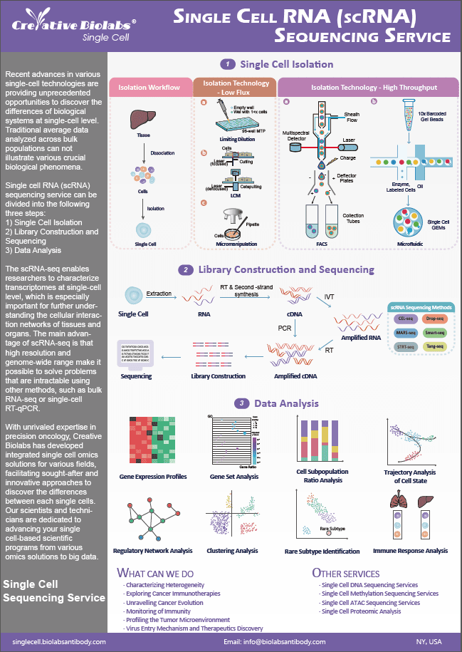
LCM is a single cell isolation method. In LCM sorting, cells do not need to be tagged by the specific antibody used for the genetic manipulation of the organism. Instead, LCM is based on selecting cells through the microscope from a part of the tissue.
Single cell isolation by LCM comprises three steps:
- A tissue is observed under a microscope and identified.
- The desired section is marked by drawing a line around it.
- A laser beam cuts the tissue along this trajectory and isolates the cells.
Then cells are transferred to the polymer film, and laser pulses activate this transfer film. Next, an infrared laser beam is used to heat the cells, which then bind to the transfer film by melting. Finally, the cells with the same morphology with intact nucleic acids and proteins are kept on the transfer film.
Advantages of LCM in Single-Cell Isolation
- It avoids any intricate operator-dependent step.
- The laser can ablate the adjacent rim of unwanted tissue, so non-specific adherence of tissue to the cap is avoided.
Limitations of LCM in Single-Cell Isolation
- Low throughput.
- Requires high skill levels.
- It can be contaminated by neighboring cells.
At Creative Biolabs, we provide tailored LCM service for a better solution for single-cell isolation. If you have any requirements, please feel free to contact us for further communication about your project.
Q&As
Q: How is the quality of samples ensured in Laser Capture Microdissection?
A: Quality control in LCM involves using high-quality tissue sections, optimizing laser settings, and ensuring minimal sample loss during the capture process. Proper staining and sectioning techniques, along with careful handling, are critical to obtaining pure and intact samples for analysis.
Q: What types of samples can be processed using LCM?
A: LCM is versatile and can be used with a variety of sample types, including fresh, frozen, and formalin-fixed paraffin-embedded (FFPE) tissues. This flexibility allows researchers to work with archival samples, expanding the possibilities for longitudinal studies and retrospective analyses in medical research.
Q: How precise is LCM in isolating individual cells from complex tissues?
A: While LCM typically requires fixed and stained tissues, ongoing developments are exploring its compatibility with live-cell imaging. This would allow researchers to observe cellular dynamics in real-time before isolation, providing a dynamic view of cellular processes prior to analysis.
Q: What expertise is required to perform Laser Capture Microdissection?
A: Performing LCM requires expertise in histology, microscopy, and molecular biology. Operators need to be skilled in preparing high-quality tissue sections, optimizing laser settings, and handling delicate samples. Collaborating with experienced service providers can help researchers access the necessary expertise.
Q: Can Laser Capture Microdissection be combined with other techniques?
A: Yes, LCM can be combined with various downstream techniques such as PCR, RNA sequencing, proteomics, and metabolomics. This integration allows for comprehensive analysis of the isolated cells or tissue regions, providing a deeper understanding of their molecular characteristics and functions.
Resources
Search...


